Everything About Knee Replacement

THE KNEE JOINT

Joints are the areas where bones meet, and movement occurs. The knee joint is made up of the femur above and the tibia below. The two bones are separated by cartilage that acts as a cushion and allows movement.

REASONS FOR SURGERY

The reasons for total knee replacement surgery in Delhi are: severe pain, loss of mobility, or deformity of the knee. Symptoms may be due to osteoarthritis, rheumatoid arthritis, or trauma among others.

Osteoarthritis, commonly called “wear and tear,” is the most common cause for a total knee replacement.

ABOUT KNEE REPLACEMENT

The knee joint is made up of the ends of the thigh bone (femur) and the shin bone (tibia). These bones normally slide over each other with ease because they are covered by soft cartilage. If an injury damages the cartilage or is worn away by arthritis, for example, it can make the joint ache or stiff.

Generally, a new knee joint improves mobility and decreases pain, although your new knee will not be able to bend as much as a normal knee joint.

Depending on the condition of your knee joint, they will replace part or all of your knee joint. A total knee replacement is more common.

Artificial knee pieces can be made of metal and / or plastic, and a knee replacement can last up to 20 years.

WHAT ARE THE ALTERNATIVES?

Knee replacement surgery in Delhi is generally recommended only if nonsurgical treatments, such as physical therapy and exercise, taking medication, or using physical support devices such as a cane, no longer help decrease pain or improve mobility.

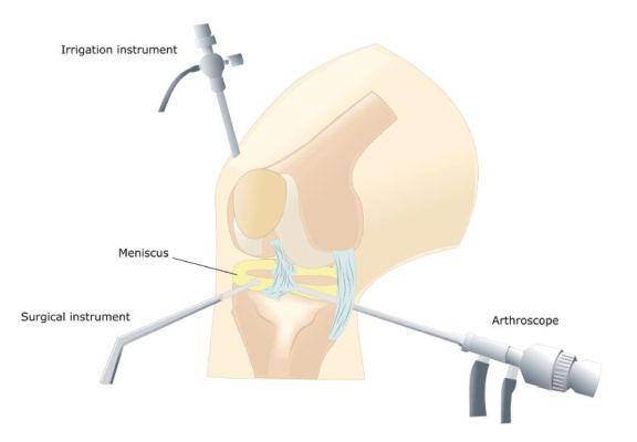
Alternative surgical procedures include arthroscopy in Delhi (if the arthritis is not very severe) or osteotomy (in which the leg bones are cut and put back). You may have already had these procedures before your knee replacement.

The surgeon will explain your options.

PREPARING FOR A KNEE REPLACEMENT

The orthopaedic surgeon in Delhi will explain how to prepare for the operation. For example, if you smoke, they will ask you to stop smoking, as this increases your risk of chest and wound infection, which can delay your recovery.

Typically, you must stay in the hospital for about five days, and the surgery is performed under general anesthesia. This means that you will be asleep during the operation. Otherwise, if you prefer, the surgery can be performed under epidural or spinal anesthesia. This type of anesthesia completely numbs from the waist down, and you will remain awake during the operation.

If you are going to have general anesthesia, you will be asked to fast. This means that you should not eat or drink, normally, for about six hours before general anesthesia. However, it is important to follow the instructions of your anesthetist.

In the hospital, the nurse can check your heart rate and blood pressure and do a urine test.
Your surgeon will explain to you what will happen before, during, and after the procedure, and any pain you may have. This is your opportunity to understand what will happen, and it may be helpful to prepare questions about the risks, benefits, and other alternatives to the procedure. This will help you stay informed so that you can give your consent if you are asked to sign a consent form to carry out the procedure.

You may be asked to wear compression stockings on your unaffected leg to prevent blood clots from forming in your veins (deep vein thrombosis, DVT). You may need an injection of a blood-thinning medicine called heparin in addition to, or instead of wearing, compression stockings.

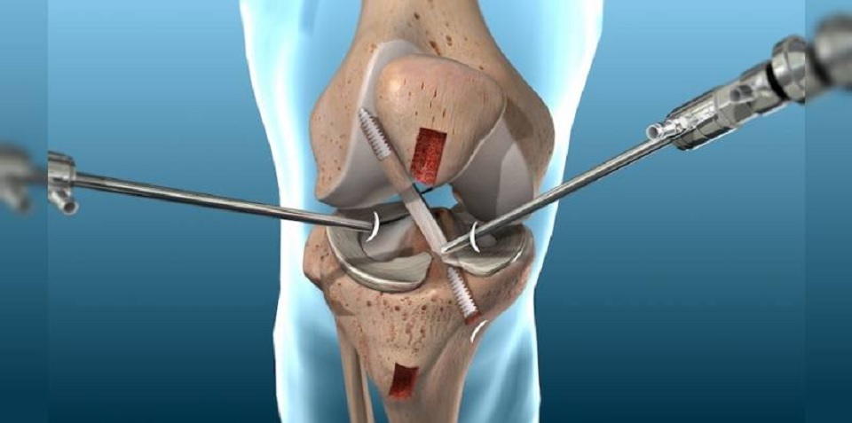
WHAT HAPPENS DURING A KNEE REPLACEMENT?

Generally, a knee replacement in Delhi takes about two hours.

The orthopaedic in Delhi will make a single cut (10 to 30 cm long) in the front of your knee. You will push the kneecap to the side to reach the knee joint. The surgeon will remove the worn or damaged surfaces from the end of the femur and the top of the tibia. Typically, he will remove the anterior cruciate ligament and may remove the posterior cruciate ligament. For support, the best orthopaedic in Dwarka will not remove the collateral ligaments. It will shape the surfaces of the femur and tibia to fit the artificial knee joint and then fit the new joint over both bones.

Sometimes the back of the kneecap is replaced with a piece of plastic. This is known as patella lining.
After placing the new joint, the surgeon will close the wound with stitches or clips and cover it with a bandage. The surgeon will place a tight bandage on your knee to help minimize swelling.

WHAT SHOULD I EXPECT AFTER

You will need to rest until the anesthesia wears off. After epidural anesthesia, you may not be able to feel or move your legs for several hours.

You may need pain relievers to ease any discomfort when the anesthesia wears off.

You may have an intermittent compression pump attached to special pads on your lower legs for the first day or so. By inflating the cushions, the pump encourages healthy blood circulation and helps prevent a DVT. You can also have a compression stocking on your unaffected leg. This helps maintain circulation.

A physiotherapist in Dwarka (a movement and mobility specialist) will visit you daily to guide you through exercises that will help you recover.

You will stay in the hospital until you can walk safely with the help of a cane or crutch. When you can go home, you will need to ask someone to drive you.

Before you go home, the nurse will give you recommendations for caring for your knee and a date for your follow-up appointment.

How long it takes for the sutures to disappear will depend on the type used in the surgery. However, for this procedure they usually go away in about six weeks. Nonabsorbable sutures and clips are removed 10-14 days after surgery.

RECOVERING FROM KNEE REPLACEMENT SURGERY

If necessary, you can take an over-the-counter pain reliever, for example acetaminophen or ibuprofen. Follow the instructions in the patient information leaflet that comes with your medicine, and if you have questions, ask your pharmacist.

Physical therapy exercises are an indispensable part of your recovery, so it is essential that you continue to do them for at least two months.

You will be able to move around your house and go up and down stairs. For a few weeks, some everyday activities, such as shopping, will be difficult for you to do. You may need to use a cane or crutches for about six weeks.

You may be asked to wear compression stockings at home for several weeks.

When resting, raise your leg and support your knee to help prevent leg and ankle swelling.

Depending on the type of work you do, you may be able to go back to work after six to 12 weeks.

Follow your surgeon’s recommendations for driving. You should not drive until you are sure that you can brake in an emergency without discomfort.

WHAT ARE THE RISKS?

Knee replacement surgery in West Delhi is a common and generally safe procedure. However, in order to make an informed decision and consent, you must be aware of the possible side effects and risk of complications associated with this procedure.

Side effects

These are the unwanted, though mostly temporary, effects of successful treatment; for example, feeling dizzy as a result of general anesthesia.

Your knee will hurt and be swollen for up to six months.

You will have a scar on the front of your knee. You may not have sensation in the skin around the scar. This may be permanent, but it should get better in two years.

Complications

Complications are problems that occur during or after the operation. Most of the people are not affected. Possible complications from any operation include unexpected reactions to anesthesia, excessive bleeding, or clot formation, usually in a vein in the leg (DVT).

Complications specific to knee replacement are rare, but include:

  • Wound or joint infection Antibiotics are given during and after surgery to prevent this complication.
  • Unstable joint. The knee joint may loosen and may require surgery to correct it.
  • Damage to blood vessels or nerves. It is usually mild and temporary.
  • Scar tissue. Scar tissue formation can limit movement. You will likely need another surgery to correct it.

Knee Arthroscopy For Knee Injuries

What is knee arthroscopy?

Knee arthroscopy is a minimally invasive procedure that allows access and treatment of injuries that affect the various structures of said joint. For this, 2 or 3 small incisions of less than one centimeter are made that allow access to the joint.

Most knee surgeries that do not involve the placement of a prosthesis are performed with a complete or partial approach with knee arthroscopy in Delhi. It is the technique of choice to address many injuries because it allows a better and greater visualization of the joint. In a non-aggressive way, all the points of the knee can be accessed to clean the cartilage, small perforations of the bone that lacks cartilage (microfractures), stabilize the cartilage and apply substances or elements that allow the cartilage to regenerate (plasma rich in platelets or stem cells).

Likewise, knee arthroscopy is also used as support for other open techniques, since it improves the diagnosis and prognosis of the patient’s injury, as it is less aggressive.

The arthroscopy in Delhi can be performed under local, regional or general anesthesia, depending on the injury and the patient. The anaesthesiologist will decide the best method for the patient, as long as he suffers as little as possible.

Why is it done?

Knee arthroscopy is used to solve knee injuries. Thus, meniscus injuries are one of the most common pathologies and, thanks to arthroscopy, it is possible to preserve most of the menisci, since the resection is not complete but partial. Meniscal sutures and the possibility of transplanting the meniscus with knee arthroscopy are common techniques that allow better protection of the cartilage of the joint.

Another of the most dangerous injuries related to sports practice is the rupture of the anterior cruciate ligament. If this is injured, it causes instability in the knee that makes it impossible for the patient to perform practically any sport. If the instability continues, it can damage surrounding structures, such as the menisci and cartilage. Hence, it is necessary to reconstruct the anterior cruciate ligament with grafts, accessing the joint through arthroscopy.

On the other hand, cartilage injuries (chondroplasties, osteoarthritis or osteochondritis) are also very common. Preserving the cartilage will also preserve the joint, avoiding wear and tear on the knee.

What does it consist of?

The orthopaedic in Delhi will make the small incisions in the knee to be able to access it. He will first fill the knee joint with a sterile solution and remove any cloudy fluid. This way he will be able to see the joint clearly and in detail.

The orthopaedic in Dwarka will then insert the arthroscope (a very thin device with a camera on the end) into the knee. This device sends the images to the television monitor, so that the surgeon can see all the structures in detail. Through the other holes, the surgeon will introduce the surgical material that will allow him to address the injury and repair the damaged structures.

It is a procedure that usually does not last more than an hour. After that, the patient will be transferred to a rehabilitation room and will be able to leave the hospital after two hours, more or less.

Preparation for knee arthroscopy

Before surgery, the patient must undergo a complete physical examination so that the specialist can assess their health and any anomaly that may interfere with the arthroscopy. Likewise, the patient must inform the orthopaedic surgeon in Delhi of the medication he takes, so that he can tell him which ones he should stop taking before the intervention. Some complementary preoperative tests will also be carried out, such as magnetic resonance imaging, electrocardiogram or blood tests.

Care after the intervention

Recovery after arthroscopy is faster than conventional open surgery. However, the advice of the specialist must be followed so that the knee recovers correctly.

It is normal for the patient to experience swelling in the days after the intervention, so it is recommended that the leg be elevated during those first days after the surgery. Also, the application of ice will relieve pain and reduce inflammation.

The incisions should also be healed, keeping them clean and dry. The orthopaedic in West Delhi will tell the patient when they can shower or change the bandage.

On the other hand, shortly after the intervention, the patient must begin rehabilitation exercises with a Physiotherapist in Dwarka, who will establish a program appropriate to the patient and the injury. This will help restore motion and strengthen your knee muscles.

Alternatives to this treatment

The alternative to knee arthroscopy in West Delhi will be conventional open surgery, which is currently only used in more serious cases, in which a prosthesis must be placed. Any other technique will suppose a greater invasion in the knee and a worse postoperative period.

All About Knee Arthroscopy

What is knee arthroscopy?

Arthroscopy in Delhi is a surgical technique that allows you to directly see the inside of the knee joint and work inside it, without having to open it. Only two small incisions or cuts are made in the skin, about one centimetre each (which is why it is called a mini-invasive technique).

Arthroscopy in Delhi is considered the best current technique for meniscal injuries, adhesions, plica, loose bodies, cartilage injuries (chondroplasty) and reconstruction of cruciate ligaments, explains the orthopaedic in Delhi.

How is knee arthroscopy done?

The orthopaedic surgeon in Delhi, in order to see the inside of the joint well and avoid tissue injury with his manoeuvres, fills it with sterile pressure serum, which has the effect of inflating a balloon; and at the same time, it allows continuous joint washing, eliminating blood residues, excised tissue fragments, etc.

The patient lies on his back on the operating table. No system is necessary to pull the joint. You only need to lock the position of the thigh and the surgeon or assistant mobilizes the leg, opening the joint space.

Through an incision, a micro camera is introduced that illuminates and amplifies the interior of the joint, viewing the image on a television monitor. On the other hand, work instruments are introduced, such as probes, hand grippers and motorized smoothing devices.

The anaesthesia used is spinal anaesthesia (patient conscious but asleep from the waist down). Some sedation may be associated with this procedure to be calmer during the surgical act. General anaesthesia is reserved for special cases.

A tourniquet is used on the thigh to prevent bleeding from the knee during the operation, thus promoting vision through the camera.

Although it is a surgical act and requires the same aseptic conditions (cleanliness and sterility to avoid infection) as any other operation, the hospital stay is usually very short. In most cases, the patient can be discharged on the same day, when the anaesthetic effect has worn off. These operations can therefore be included in the program of major outpatient surgery, explains the orthopaedic surgeon in Delhi.

What does knee arthroscopy in Delhi show?

  • The appearance of synovialfluid (viscous fluid that lubricates the joint), which may be cloudy, contain blood or loose bodies, usually cartilage. Synovial fluid can be analysed to determine its composition in special cases.
    • The synovial membrane(the sac that lines the joint inside and produces synovial fluid). In certain cases, a sample (biopsy) is taken for analysis under a microscope.
    • The cartilage that lines the articular surfaces of the femur, tibia, and patella. It is palpated with a special hook to see the consistency and it is observed if it has injuries: wear (osteoarthritis), fissures, chondromalacia …
    • The menisci (internal and external): observed and palpated with the probe hook. Breaks, tearing, wear are detected …
  • The cruciate ligaments(anterior and posterior): they are seen and touched to determine partial or total tears, laxity, function … The collateral ligaments are not seen with this technique.
    • The way the patellamoves when the knee is bent and stretched, as well as the friction surfaces.

In which cases should an arthroscopy in Dwarka be performed?

Less and less to diagnose, as advances in ultrasound, CT (scanner) and nuclear magnetic resonance resolve it more and more frequently, although they are not infallible. 

However, in cases of doubt or when a major intervention on the knee is planned, an arthroscopy can be performed beforehand, which will make it possible to confirm the diagnosis, rule out other injuries and decide the best possible treatment, which is also sometimes arthroscopic. Thus, in the same surgical act it is diagnosed and treated. In addition, there are patients who have contraindicated MRI (due to claustrophobia, or prosthetic heart valves), in those cases, diagnostic and therapeutic arthroscopy would be indicated by the orthopaedic doctor in Delhi.

Another diagnostic utility is to allow a synovial biopsy in certain diseases.

Currently, the main indications for performing a knee arthroscopy in Dwarka are:

  • Meniscal injuries: remove broken fragments, suture certain tears, regeneration techniques and meniscal reimplantation
  • cruciate ligament reconstruction: avoid opening the knee as before
  • cartilage injuries: cleaning, regenerative techniques (platelet growth factors, mosaicplasty)
  • removal of intra-articular loose bodies: fragments of detached cartilage or meniscus
  • removal of synovial plica or synovitis (synovial membrane hypergrowth)
  • cleaning on knees with osteoarthritis (wear) before reaching the total knee replacement

Recommendations at hospital discharge:
They are usually quite simple since it is a mini-invasive technique. 

A compression bandage is placed, which the patient will remove at home after 48 hours. Then the first treatment is carried out, which consists of painting the two small wounds with Betadine and covering them with two adhesive dressings. 

From there, the treatment will be repeated every day until the stitches fall out (about 2 to 4 weeks). It can be made to coincide with the shower as long as two rules are met:

  • quick shower: the less time the wounds are wet the better
  • WITH the dressings on: so that soap, shampoo, dirty water do not get into the wounds

After showering, the wet dressings are removed, the wounds are thoroughly dried with sterile gauze, painted with Betadine, and new dressings are placed.

From the moment the mobility of the legs recovers after anaesthesia, it is advisable to start walking. At first helped with crutches and following the indications of your orthopaedic surgeon in Dwarka regarding load (partial or complete). Usually, the patient leaves the hospital the same day walking with the help of two crutches.

An anti-inflammatory treatment is usually recommended at discharge for the first days.

It is advisable to apply ice locally for 10-15 minutes about 3-4 times a day to help reduce inflammation.

Depending on the diagnosis and treatment carried out, a specific physiotherapy may or may not be prescribed, with the recovery times greatly varying. The time in which you can return to sports or hard work depends on the injury: from 1 month to several months.

In the event of residual effusion (usually due to the persistence of the arthroscopic lavage fluid and more rarely due to bleeding into the joint, which is the hemarthros), an evacuating puncture may be necessary: ​​the area is punctured, and the excess fluid is extracted with a syringe. This procedure should only be performed by an orthopaedic surgeon in Delhi.

Knee arthroscopy: What are its benefits?

Thanks to knee arthroscopy, the orthopaedic surgeon in Delhi is able to see the joint completely while operating. Find out more below.

Knee arthroscopy in Delhi is a surgical procedure used in traumatology and orthopaedic surgery. This technique facilitates diagnosis and treats many problems and diseases located inside the joint.

In different part of world, it has been commonly used since the 1970s. For this reason, despite being carried out for years, it is still considered as a new technique in continuous advancement and expansion.

Keep in mind that, despite being a much less invasive method than others, knee arthroscopy in Delhi is a surgical procedure. For this reason, only surgeons specializing in traumatology and surgery should perform it.

The image it offers is collected thanks to a camera and only a minimal incision that barely leaves a scar is necessary. It is a non-invasive method that allows to reduce hospital admission times. In most cases, patients are discharged on the same day, facilitating recovery, explains the orthopaedic doctor in Delhi.

Indications of knee arthroscopy

This technique allows to visualize the inside of the knee in a minimally invasive way.

There are several problems or injuries in which this useful technique is used. Diseases of various types and injuries can damage different parts of the joint such as:

  • bone.
  • cartilage.
  • Meniscus.
  • tendons.
  • muscles.
  • ligaments.

Ligament injury

Cruciate ligament injury is one of the injuries that require knee arthroscopy in Dwarka for accurate diagnosis. The injury can occur in both the anterior and posterior ligaments.

The anterior cruciate ligament is the isolated ligament with an important stability function. The name is because it crosses with another ligament called the posterior cruciate ligament, which has an antero-posterior and lateral half direction.

This injury is the most common intraarticular problem and is usually caused by lateral leg twisting. In addition, if associated with internal meniscus rupture and internal lateral ligament, it is called triad. It is very common in footballers, says the sports injury specialist in west Delhi.

In a young person, moderately active, with discomfort and feeling of failure, the ligament must be reconstructed. This is because in the medium term it usually causes early degeneration of the joint and predisposes to meniscus injury, explains the orthopaedic in Delhi.

Meniscal injury

Meniscus injuries are often one of the main indications for using this technique, both in diagnosis and repair.

Meniscus is a cartilage that has important functions within the knee such as:

  • cushioning.
  • Cartilage protection.
  • Improve joint function.

Sometimes, when a clean, recent and peripheral injury occurs, the meniscus can be sutured. There are two types of suture: all with a knee arthroscopy, or mixed, less frequent.

With a knee arthroscopy in Dwarka, meniscus transplants from tissue donors can also be performed.

Wear or degeneration of joint cartilage.

Cartilage is a pearly layer of low friction that coats the joint part of the femur, tibia and kneecap. The most common cause of cartilage lesions is osteoarthritis.

However, more triggers are in this situation. It can also be caused by osteochondritis desiccant, infections, metabolic problems or trauma, among others. Depending on the patient’s age, activity and expectations, there are several surgical techniques for repairing or reconstructing cartilage.

Benefits of knee arthroscopy in Delhi

Thanks to this technique the hospitalization time is reduced, facilitating a prompt recovery of the patient.

The advantage of this technique over other common surgical procedures is that the orthopaedic surgeon in Delhi is able to see the joint completely. It does this thanks to a small instrument called an arthroscope.

Another advantage of knee arthroscopy in Delhi is that only very small incisions are needed. Thus, usually this technique leads to a reduction of the time of stay in the hospital and a rapid recovery.

Many patients who have knee arthroscopy can leave the medical centre the same day they are operated on. In turn, being small incisions, a better aesthetic result is achieved, especially in exposed areas of the body.

But don’t forget that patients undergoing knee arthroscopy can do so due to different injuries or pathologies. Therefore, the previous particular conditions will condition the stay in the hospital and the total recovery time. In short, not all patients will respond equally to this surgery, says the orthopaedic in Delhi.

Shoulder Bursitis – What it is, Symptoms, Treatment, Cure

What is shoulder bursitis?

The shoulder bursitis results from an inflammation of the synovial bags (or bursae) that exist around this joint and the tendons of the rotator hood.

It is one of the most frequent causes of pain in this joint. It can be bilateral, reaching both the left shoulder and the right shoulder.

It can be acute or progress to chronic shoulder bursitis, says the orthopaedic in Delhi.

Subacromial bursitis

There are several bursitis that can occur in the shoulder depending on whether one or the other synovial pouch is affected.

The most frequent are those that reach the sub deltoidal subacromial bursa, forming what is commonly called subacromial bursitis.

Shoulder bursitis – symptoms

Shoulder bursitis presents a clinical picture similar to that of tendonitis in the shoulder.

The most characteristic symptoms are the presence of inflammatory pain, located on the antero-lateral face of the shoulder, eventually radiating to the arm and elbow. Its worsening is especially felt with efforts or during the night, making it impossible for the patient to sleep on the affected shoulder.

Also characteristic of shoulder bursitis is the presence of crackling that is palpable, or perceived by the patient, when mobilizing the joint, explains the orthopaedic doctor in Delhi.

Shoulder bursitis – causes

The most frequent causes of shoulder bursitis are trauma and repeated efforts, such as those that occur with certain work activities (painters or plasterers, warehouse replenishers, etc.) or with the practice of certain sports, such as weight training, swimming or others practiced with the arm above the head (“overhead sports”), says the sports injury specialist in west Delhi.

Certain rheumatic diseases (such as, for example, rheumatoid arthritis, gout, lupus, psoriatic arthritis) also often develop with bursitis (namely subacromial).

Shoulder bursitis – diagnosis

The diagnosis of this pathology is made, essentially, through a careful clinical examination, carried out by your specialist shoulder orthopaedic doctor in Delhi.

The exams to be performed later may be an x-ray that helps to exclude other causes of shoulder pain (such as osteoarthritis or calcifying tendinitis) and an ultrasound of the shoulder that easily visualizes the fluid in the subacromial bursa (caused by the inflammatory effusion).

The nuclear magnetic resonance (NMR) of the shoulder is, however, one that has a better diagnostic accuracy, obtaining high-resolution images of the surrounding muscle-tendon structures and eluding any intra-articular pathology, states the orthopaedic in West Delhi.

Is shoulder bursitis curable?

Shoulder bursitis is curable. Next, learn how to treat shoulder bursitis.

Shoulder bursitis – treatment

The medication or remedy most commonly used to treat shoulder bursitis is anti-inflammatory. The most common are ibuprofen, diclofenac, naproxen, among others. Its use is aimed at relieving pain and decreasing the inflammation that is always present in these conditions. Its application can be topical in the form of creams (or gels), or impregnated dressings that will make a prolonged release of these agents in the affected area.

The patient can also take these drugs in oral form (in capsules or diluted), taking care to do so after meals or preceded by taking gastric protectors, in order to avoid the aggression of the gastric or duodenal mucous membranes, states the orthopaedic doctor in Dwarka.

The physiotherapy is essential to maintain joint function and help control pain. A well-oriented physiotherapeutic treatment, with adequate exercises, can prevent the installation of a marked stiffness in the shoulder, usually called adhesive or retractable capsulitis (frozen shoulder or “frozen shoulder”). This would oblige to prolong the treatment for its complete resolution.

In cases of very acute and incapacitating pain, or reluctant to any other non-invasive treatment, infiltration or local injection of corticosteroids diluted with local anesthetic, may be a therapeutic weapon to be used by your orthopaedic doctor in Dwarka to resolve the condition. Your institution should be judicious and always consider the pros and risks of its application.

Shoulder bursitis – surgery

Shoulder arthroscopy in Delhi (surgery) is reserved for cases that are resistant to medical treatment or that simultaneously present other pathologies, such as rotator cuff tears or osteoarthritis, says the orthopaedic surgeon in Delhi.

This operation is performed using mini-invasive techniques that allow an excellent articular visualization, performing a minimal surgical aggression and thus providing a well-tolerated postoperative period and an easier and faster rehabilitation.

It should be carried out in specialized orthopaedic clinic in Delhi and by experienced orthopaedic surgeon in Delhi in order to obtain the best results.

Elbow Dislocation. Causes and types of treatment

What is an elbow dislocation?

Elbow dislocation is a very common pathology in traumatology. This occurs when the relationship between humerus, ulna and radius is broken, and the pain that it produces in the person who suffers it is very intense, says the orthopaedic in Delhi.

When the articular surfaces of an elbow are separated, the elbow is displaced. Elbow displacement can be complete or partial. In complete displacement, the joint surfaces are completely separated. In a partial displacement, the surfaces of the joints are only partially separated. A partial displacement is also called a subluxation.

For this reason, we suggest that you consult an orthopaedic doctor in Delhi specialized in arthroplasty and elbow arthroscopy in Delhi, techniques through which elbow injuries can be solved quickly and in a minimally invasive way.

It is important to note that expectations in the case of this injury are very high and recovery occurs satisfactorily in at least 80% of cases.

What are the causes of elbow dislocation?

The main reason for this injury is a heavy fall on the outstretched hand since there is a high chance that the humerus will be out of its correct alignment with the joint, explains the orthopaedic doctor in Delhi.

Traffic accidents are also a common cause due to severe trauma.

Elbow dislocation can occur in sports such as soccer, hockey, skiing, gymnastics, or volleyball.

In this sense, sports practice becomes one of the risk factors along with age (children’s elbows are more prone, so we must avoid jerks when walking), sex (affects more men than women) or heredity (people with looser ligaments).

How is the preoperative elbow dislocation?

First of all, the bones must return to their place and for this an orthopaedic in Delhi will perform a reduction, but not before correctly evaluating the injury so as not to cause further damage. Subsequently, the pathology will be studied to decide whether to perform an elbow surgery that will be necessary in the following cases:

  • Fracture of any of the dislocated bones.
  • Ligament tear.
  • Damage to nerves or blood vessels and bones of the elbow.

In the case of operating on the patient, the team of orthopaedic surgeon in Delhi prepares thoroughly for possible complications with a set of fundamental tools in the operating room:

  • Radius Head Fracture Fixation Kit, Coronids, Proximal Ulna.
  • Implants to replace radio head.
  • Bone anchors for soft tissues.
  • Articulated external fixation

What are the types of elbow dislocation treatments?

Orthopaedic surgeon in Delhi proposes two possibilities to treat elbow dislocation that we expose below and that will be useful one or the other according to each case:

Acute treatment:

It is applied in cases where there are fractures associated with the head of the radius and / or coronoid and the approach can be lateral or medial. Specifically, this surgery is used to successfully repair radial head fracture, proximal ulna and coronoid tear, and lateral ligament.

Likewise, in this type of treatment articulated external fixators are sometimes used for the following cases:

  • Neutralization of the load on the repairs when they are seems weak.
  • The elbow appears vulnerable to reluxation.
  • So that the patient can move the arm from the first day in a regulated way.

Deferred treatment:

The parts that are restored are the same as with the acute treatment but with complications such as scar tissue, partially or totally united bone fragments, little identifiable joint surfaces.

If you have suffered an elbow dislocation and need advice, do not hesitate to contact orthopaedic in Delhi to assess your case, establish the appropriate diagnosis and carry out your recovery with optimal results.

Hip Arthroscopy- All You Need to Know

This was probably the area of ​​Hip Surgery that presented the biggest and most expressive evolution in the last years.

The term arthroscopy in Delhi refers to a surgical technique, the act of “looking at the joint”. It is a method, a means of carrying out a treatment. It allows different procedures to be performed on or near the joint.

This is possible through the use of micro-cameras and special instruments, of small caliber, that give us access to the interior of the joints without the need for surgical accesses of traditional surgeries.

In arthroscopy, we use cutaneous incisions of about one centimeter each, in varying numbers – usually from 2 to 4 accessions, explains the orthopaedic in Delhi.

What can be treated by hip arthroscopy in Delhi?

The most frequent indications are:

  • Femoral-acetabular impact: for bone and cartilage remodeling (osteochondroplasty).
  • Lesions of the acetabular Labrum: for its resection or reinsertion to the bone bed with the use of anchors.

Other indications include:

  • Snap in hip.
  • Removal of free bodies or foreign bodies from the joint.
  • Repair of injuries to the gluteal tendons and lateral hip pain syndrome.
  • Treatment of traumatic and atraumatic cartilage lesions.
  • Lesions of the round ligament.
  • Osteochondritis dissecans.
  • Synovectomy: for rheumatoid arthritis or villonodular synovitis.
  • Deep gluteal pain syndrome or piriformis syndrome: to release the sciatic nerve.
  • Some sequelae of pediatric pathologies, such as Legg-Perthes: for removal of free bodies or chondroplasty.
  •  

Can my case be treated by arthroscopy in Delhi?

Through clinical evaluation and imaging tests we can define who will benefit or not from this type of treatment.

Although this surgery is extremely versatile, not all hip injuries can be treated in this way.

Cases of major morphological changes, hip dysplasia and osteoarthritis, for example, are likely to be better treated through conventional surgery, when indicated.

Although common sense is that arthroscopy in Dwarka is “better and more modern”, this is not always true. Each case has its indication. In some cases, traditional surgery may bring more benefits.

The indication of treatment must be decided on a case-by-case basis by the orthopaedic doctor in Delhi.

How is the surgery?

Surgery can be performed with spinal anesthesia and sedation or under general anesthesia.

To gain access to the hip joint, it is necessary to use an orthopedic traction table, under anesthesia, creating space in the joint for the insertion of optics and instruments.

In addition to the video material, we also use a radioscopy device, which is a kind of “live” radiography.

After making the access portals, we introduced the micro-camera and we can use a wide variety of tweezers to correct injuries, sutures, scrapes, micro-perforations, etc.

The instruments used in hip arthroscopy are specific to this surgery, being longer than the material traditionally used for knee and shoulder, for example.

We also have flexible instruments that can deviate from the femoral head and increase our range within the joint, however, the shape and location of the hip imposes a natural limit on the range of the instruments, explains the orthopaedic surgeon in Delhi.

What are the risks?

As with any surgical procedure, there are risks involved.

In addition to the “general” risks, such as: anesthetic risk and infection, there are also specific risks of arthroscopic treatment, especially paresthesias (alteration of sensitivity) in the inguinal region or skin problems, among others. The evolution of technique and materials has fortunately made these problems increasingly rare.

In addition, as with all endoscopic surgery, there may always be a need for conversion to traditional surgery (“open air”).

How is recovery after surgery?

According to the severity of the injury and the treatment instituted, specific physiotherapy protocols are initiated. The use of crutches is necessary for two to six weeks and the return to sports is allowed according to the type of injury, the type of sport and the individual recovery. Final recovery can take anywhere from six months to a year, states the orthopaedic surgeon in Delhi.

It is usually necessary to stay in the hospital for a day for post-anesthetic recovery and the postoperative period is generally not very painful.

Will I never have hip pain again? Can I go back to sports?

Statistically up to 80% of patients are able to resume their sports activities with at least partial relief of symptoms.

In practice, each case is different. Depending on the problem you had in your hip, arthroscopy can only be palliative, in an attempt to preserve your joint. In some situations, it may be more prudent to abandon impact sports altogether.

Eventually the symptoms may remain or appear again after a while. You should inquire about the details of your case with your orthopaedic surgeon in Dwarka.

In our practice, we observed the best results in those patients who obtained an earlier diagnosis and in small lesions, without compromising cartilage.

For this reason, you should not neglect your symptoms, in case of pain in the groin or hip always look for an orthopaedic in Delhi.

What is an Arthroscopy and its Advantages?

Arthroscopy in Delhi is a minimally invasive surgery, which is used for the diagnosis and treatment of the joints. It is performed by introducing an arthroscope connected to a high-resolution camera, which allows the joint to be viewed in its entirety through a high-definition monitor.

This technique has many benefits and is increasingly being used to treat joint diseases. It allows a more complete view of the joint, through minimal incisions, which are made under local anesthesia. As it is a minimally invasive intervention, it offers good results and the patient’s recovery times are reduced, states the orthopaedic in Delhi.

Types of arthroscopy

It could be said that there are no different types of arthroscopy in Delhi, since the technique used is always the same, however, the differences arise depending on the joint operated.

The most common arthroscopies are the following:

  • Shoulder
  • Elbow
  • Doll
  • Hip
  • Knee
  • Ankle

When is an arthroscopic operation performed?

Arthroscopy in Delhi is a technique that is carried out to visualize the inside of the patient’s joint. In many cases, it is carried out as a diagnostic test, for example, to take samples or perform biopsies of the area, in case of tumors or intra-articular lesions.

However, in other cases, it is carried out for the treatment of joint injury, for example, to remove cysts, perform intra-articular washes or remove extra-articular bodies that can cause inflammation of the patient’s joint. It is important that you are always advised by a specialist orthopaedic doctor in Delhi.

How is the arthroscopic operation?

Arthroscopic intervention is usually carried out using local anesthesia at orthopaedic clinic in Delhi, that is, localized anesthesia in the area to be intervened. First, a minimal incision is made by the orthopaedic surgeon in Delhi, through which the arthroscope will be inserted.

Once the incision has been made, cartilaginous cleaning is carried out, that is, debridements are corrected in order to provide stability to the cartilage. In many cases, a treatment with plasma rich in platelets or stem cells is applied, which help the regeneration of the intervened tissues, explains the orthopaedic in Delhi.

Advantages of arthroscopy

Arthroscopic interventions have many advantages over traditional surgeries. First of all, it is a minimally invasive technique, the incisions are of a minimum size, which makes the risk of suffering infections considerably lower.

In addition, the patient recovers quickly and the time it takes to be able to return to their normal activities is reduced, says the orthopaedic in Delhi.

How is recovery after arthroscopy?

As explained, the recovery after an arthroscopic operation is fast, since the invasion into the tissues is minimal. Proper care of the incisions is especially important to avoid infections that can affect recovery.

Usually, the patient will start a rehabilitative treatment one month after the operation, with the aim of strengthening and restoring joint mobility. Three or four months after surgery, the patient will be able to recover his usual activities, avoiding activities that imply an impact on the joint, explains the orthopaedic in Delhi.

Rehabilitation after arthroscopy

In arthroscopic interventions, rehabilitation is of particular importance, which can be carried out under the supervision of an orthopaedic doctor in Dwarka or a specialized physiotherapist in Dwarka. The objective is to strengthen the joint, to prevent the injury from developing again, and to regain joint stability.

In this process, it is advisable to carry out sports practices that do not imply a great impact on the joint, for example, walking. In addition, the patient must perform prescribed flexion and extension exercises, which help to regain full mobility of the joint, says the orthopaedic in Dwarka.

As explained, arthroscopy in Dwarka is a technique that can correct injuries that are disabling for the patient and help him regain his quality of life.

SHOULDER PAIN WHEN RAISING AN ARM: CAUSES AND TREATMENT

The inability to raise your hand up is a fairly common problem. Shoulder pain can be one of the most tiring and painful conditions. Many of you will one day just wake up with this pain. Pain can be localized to the top or side of the shoulder joint. In some patients, pain is most pronounced in the evening and at night, in others it occurs only when the arm is raised up. Most of you will not remember any kind of shoulder injury. Most did not change their training regimes or make them more aggressive. In most cases, you are 40-60 years old, and this could be your dominant or non-dominant hand. Let’s take a look at the most common causes:

  • Pain in the upper shoulder
  • Shoulder pain when lifting the arm
  • Restriction of movement in the shoulder joint.

According to the orthopaedic in Delhi, there are many problems that can lead to limited shoulder movement or pain when lifting or moving the arm. Most often these are problems with the rotator cuff.

The rotator cuff is the most common cause of outer shoulder pain in adults. This is also the main reason why you are unable to raise your arm up or are in pain when moving, explains the orthopaedic in West Delhi.

Shoulder pain can be caused by injury. Believe it or not, shoulder injury is a less common cause of pain.

Rotator cuff injuries are more likely to occur as a result of repetitive stress, for example, when working with weights, when throwing and throwing sports equipment. Damage to the rotator cuff can also occur from falls and prolonged work with raised arms (for example, cutting trees or painting a house).

The rotator cuff of the shoulder is formed by four muscles located under the deltoid muscle. Together, these muscles control movement in the shoulder joint, ensuring normal shoulder function.

If the rotator cuff becomes damaged, inflamed, or torn, you are unable to raise your arm upward.

Let’s take a quick look at each of the most common causes of pain and limitation of movement in the shoulder joint.

ROTATOR CUFF TENDONITIS

In persons over 30, rotator cuff tendinitis is very rare, and the most common cause of pain in the shoulder joint when lifting the arm up is rotator cuff tendinosis.

The term tendinosis refers to degenerative changes in the rotator cuff. Along with degeneration in the rotator cuff tendons, the number of blood vessels and nerves increases. This is how the body responds to degeneration and this is what causes the pain. The appearance of new nerve endings leads to pain during certain movements. If the pain is severe enough, you cannot raise your arm and sleep on this side.

Physiotherapy is usually effective in patients with rotator cuff tendinosis. If, despite treatment, pain persists, surgery is possible, which helps to reverse the changes and reduce the severity of pain, says the orthopaedic surgeon in Delhi.

PARTIAL TEARS OF THE ROTATOR CUFF

As you age or degenerate, a portion of the rotator cuff may separate from the bone to which it is attached. This is a natural stage in the progression of tendinosis. When a significant part of the rotator cuff is separated, a small gap or defect appears in it. This condition is called partial rupture. Partial tears are not large enough to cause impaired movement. However, if this gap is accompanied by pain, then the pain may be localized in the upper part or along the lateral surface of the shoulder. It will also hurt to raise your hand up.

Some partial tears hurt, others don’t. You can determine this during the examination. Most patients with partial tears of the rotator cuff respond well to physiotherapy. If physical therapy or other nonsurgical treatments are ineffective, surgery is likely to help relieve night-time pain and pain when raising an arm, says orthopaedic in Dwarka.

CALCIFIC TENDONITIS

Rotator cuff calcific tendonitis is a very painful condition. It is quite simple to diagnose it precisely due to the severity of the pain syndrome. The condition is characterized by the appearance of deposits of calcium crystals in the thickness of the tendons. Fortunately, in most such cases, surgery is not needed, and it is enough to inject a drug into the focus under ultrasound guidance, which helps to wash out calcium crystals. The operation is necessary only in rare cases.

PAIN IN THE SHOULDER JOINT AFTER INJURY

Some of you may be reading this article because you fell on your shoulder and you cannot raise your hand after that. In the event of injury, there is a possibility that the rotator cuff has torn off the bone. Therefore, in such situations, we recommend that you quickly contact a specialist. Now we’ll talk about what to look out for if you suspect you have a serious shoulder injury.

The rotator cuff controls movement in the shoulder joint. If a rotator cuff rupture occurs, your complaints and symptoms will depend on the size of the rupture. For the most significant breaks, you will not be able to raise your hand at all. Many patients with fresh massive tears in the rotator cuff require surgery to restore shoulder function. Thus, treatment for a rotator cuff tear depends on the cause of the tear.

ADHESIVE CAPSULITIS OR FROZEN SHOULDER

Adhesive capsulitis or frozen shoulder is a fairly common cause of pain and limited movement in the shoulder joint. The pain in this case can be localized in the upper shoulder or axillary region. In this condition, the ligaments and capsule around the joint thicken, become tense and inflamed. Diagnosis is simple enough … by definition, frozen shoulder is a lack of movement in one shoulder joint compared to the opposite one. Treatment for frozen shoulder is usually conservative and includes physical therapy. With the help of the block system, you can independently stretch the capsule of the shoulder joint. Sometimes orthopaedic doctor in Delhi can recommend the injection of corticosteroids into the joint to control inflammation. If physical therapy proves to be ineffective, doctor may recommend going for arthroscopy in Delhi.

ARTHROSIS OF THE ACROMIOCLAVICULAR JOINT

The acromioclavicular joint (ACS) is a small joint located at the top of the shoulder. This is the joint between the clavicle and the shoulder blade. Over time, this joint can undergo degenerative changes. ACS arthrosis is the # 1 cause of pain in the upper shoulder. With sufficient severity, this pain leads to a limitation of lifting the arm up and movement in the shoulder joint. Ice, heat, and anti-inflammatory drugs can help cope with pain in such cases. It is also possible to inject drugs directly into the joint. If this treatment is ineffective, surgery is possible.

The causes of shoulder pain vary by age. Some problems are more common in certain age groups. Tendonitis is more common in athletes at a younger age. In middle age, tendinosis, calcific tendonitis, arthrosis of the acromioclavicular joint (ACS), and frozen shoulder.

Orthopaedic surgeon in Delhi widely uses arthroscopy and other minimally invasive methods of treating shoulder joint pathology. Operations are carried out on modern medical equipment from major world manufacturers.

However, it should be noted that the result of the operation depends not only on the equipment, but also on the skill and experience of the surgeon. The orthopaedic in Delhi has extensive experience in treating diseases of this localization for many years.

Knee Arthroscopy For Better Mobility of Your Knee

Knee Arthroscopy

Arthroscopy in Delhi is a surgical intervention allowing the exploration of the joint using a tube a few millimeters in diameter, introduced into the knee through several tiny holes (2 to 4) (about 1 cm). This tube, fitted with optics coupled to a miniaturized video camera, is connected to a color television screen on which the main elements of the knee will be viewed:

  • the cartilages of the femur, tibia and patella
  • the cruciate ligaments (anterior and posterior),
  • menisci (internal and external),
  • synovial membranes

ARTHROSCOPY THEREFORE ALLOWS:

  • to specify the possible causes of a knee dysfunction: pain, blockage, swelling, instability, in addition to the clinical and radiological examination, then guiding at best the choice of the subsequent treatment which will be proposed.
  • to perform a certain number of surgical procedures using fine instruments, “without opening the knee” under the control of the television screen.

THESE INTERVENTIONS CAN BE:

  • meniscal lesion removal
  • ligament plasty (cruciate ligament operations)
  • regularization of cartilage when it is irregular or worn,
  • removal of free bone or cartilage fragments in the joint,
  • synovial biopsy
  • release of adhesions,
  • fold section (plica)

IN PRACTICE

Arthroscopy in West Delhi is performed in the operating room, under anesthesia. The terms of this will be decided between you and the anesthesiologist. You will be admitted to the hospital the same morning; you must bring the x-rays as well as the biological examinations in your possession and report the usual treatments in progress.

On the day of the arthroscopic surgery in Delhi you must be on an empty stomach and respect the  instructions of orthopaedic surgeon in Delhi.

As soon as you get back to your room, you can start to gently mobilize your knee, walking being allowed with the help of the nurse or physiotherapist a few hours later.

The exit from the hospital will generally be rapid. During this, you will be given:

  • a first consultation appointment,
  • a rehabilitation order intended to improve functional recovery,
  • a work stoppage suited to the nature of your knee injuries and the type of professional activity you exercise.

During the first week, you will be advised:

  • not to bend the knee more than 90 °,
  • to walk or climb the stairs moderately,
  • avoid prolonged immobile standing, – apply an ice pack (4 times – 10 minutes a day),
  • to contract the extended quadriceps leg muscle many times,
  • not to resume sport before medical authorization

Arthroscopy is a surgical procedure.

If the hazards inherent in this technique are rare, do not hesitate to report any local incident to orthopaedic in Delhi that would worry you: persistent fever, increasing pain, hematoma or significant swelling of the knee (moderate, painless swelling is however usual with possible sensation of ” splashing “due to temporary persistence of fluid in the knee).